

Master Angular Magnification and Optical Instruments for NEET Physics
A Comprehensive Guide to Simple Magnifiers, Compound Microscopes, and Retinal Vision

This study guide is designed to help NEET aspirants master the concepts of Angular Magnification and Optical Instruments. Since visual clarity and problem-solving speed are key for the NEET exam, these notes focus on conceptual reasoning and the practical application of formulas.
Introduction
Optical Instruments: The Physics of Magnifying Power
In the study of optics, specifically for competitive exams like NEET, a clear distinction must be made between how we measure “enlargement.” While we often use the general term “magnification,” optical instruments such as microscopes and telescopes specifically utilize Angular Magnification, often referred to as Magnifying Power (M). This is fundamentally different from the Linear Magnification (m) used for mirrors and single lenses. Linear magnification is the ratio of the physical height of the image to the object (m = hi / ho). However, this measurement is only practical when an image is projected onto a screen. For instruments where the eye is the final detector, the “physical height” of a virtual image is less important than the space it occupies in our field of vision.
The human eye does not perceive the absolute size of an object; it perceives the Visual Angle (θ) subtended by the object at the eye. Angular Magnification is defined as the ratio of the angle subtended by the final image at the eye (θ′) to the angle subtended by the object when viewed directly by the naked eye (θ). For a microscope, the reference angle (θ) is measured with the object at the Near Point (25 cm), whereas for a telescope, it is the angle subtended by the distant object. By increasing this angle, the instrument “stretches” the image across a larger area of the retina, stimulating more photoreceptors and allowing the brain to resolve finer details.
The Microscope: Resolving the Microscopic
In a microscope, the primary challenge is that tiny objects subtend an angle too small for the retina to distinguish. If we attempt to increase this angle by bringing the object closer than 25 cm, the eye cannot focus, resulting in a blurred image. The microscope solves this “Focus vs. Size” conflict by using lenses to create a large visual angle (θ′) while ensuring the image is formed at a distance where the eye can focus comfortably. If we were to use Linear Magnification (m = v / u) for a microscope in “Normal Adjustment” (image at infinity), the value would mathematically be infinite. This is physically useless to a student. However, the Angular Magnification remains a finite and constant value, representing how much the “perceived” size has increased compared to the best possible naked-eye view.
The Telescope: Resolving the Distant
For a telescope, the concept of Linear Magnification (hi / ho) becomes even more problematic. Consider a distant star that is millions of kilometers wide. If a telescope creates a 10 cm image of that star, the linear magnification would be a tiny fraction, suggesting the telescope makes the star “smaller.” This is clearly incorrect in the context of observation. The telescope’s true purpose is to take an object that is physically massive but subtends a tiny angle because of its distance and expand that Visual Angle. Thus, we strictly use the ratio of the angle through the eyepiece to the angle at the naked eye. This explains why the “Magnifying Power” of a telescope is the only relevant metric for an observer.
Summary of NEET Application
Students must remember a “Golden Rule” for optical problems: Whenever a question asks for the “Magnifying Power” of an instrument, you must calculate the Angular Magnification (M). You should only use the height ratio (hi / ho) if the question specifically demands “Linear” or “Lateral” magnification.
Microscope Logic: Magnifying power is the ratio of the angle subtended by the image to the angle subtended by the object at the Near Point 25 cm.
Telescope Logic: Magnifying power is the ratio of the angle subtended by the image to the angle subtended by the distant object at the objective.
Universal Truth: In both cases, the goal is to increase the Retinal Image Size by expanding the visual angle, tricking the brain into seeing a detailed, enlarged version of reality.
NEET Physics Notes: Angular Magnification & Optical Instruments
1. Fundamentals of Vision
The human eye acts like a camera where the lens focuses light onto the retina. The clarity and size of what we see depend entirely on the Visual Angle (the angle subtended by the object at the eye).
Visual Angle (θ): Defined as the angle formed by the lines of sight from the eye to the edges of the object.
Retinal Image Size: The height of the image formed on the retina (hi) is directly proportional to the visual angle (θ).
The Logic: If you want to see a tiny object (like a bacteria) clearly, you must increase the angle (θ) it makes at your eye.
2. The Physical Significance of Angular Magnification
While linear magnification tells us how many times larger an image is compared to the object, Angular Magnification (M) tells us how much larger the image appears to “our brain”. Why the “Brain” Perceives Angles, Not Meters is as follows: -
a. The Retinal Reality
Your brain doesn’t have a ruler to measure the actual height of an object in centimeters. Instead, it counts how many light-sensing cells (photoreceptors) on the retina are being stimulated.
Physical Fact: A larger Visual Angle (θ) spreads light over a larger area of the retina.
Brain’s Interpretation: “If more of my retina is covered, the object must be larger or closer”.
b. The “Moon vs. Thumb” Paradox
Consider why you can “cover” the moon with your thumb:
Linear Size: The moon is thousands of kilometers wide; your thumb is 2 cm.
Angular Size: Because your thumb is very close, it subtends a larger angle at your eye than the distant moon.
Brain’s Perspective: Your brain sees the thumb as “bigger” in your field of view because it occupies a larger angular space.
c. Why Angle Matters More Than Size
The human eye does not perceive the “absolute size” of an object. Instead, it perceives the size of the image formed on the retina. This retinal image size is directly proportional to the Visual Angle (θ) subtended by the object at the eye.
Small Angle (θ): Results in a small image on the retina and low clarity.
Large Angle (θ’): Results in a large image on the retina and high clarity.
d. The Biological Limit (The “Why” behind M)
If we want to see a tiny object (like a cell) clearly, the natural instinct is to bring it closer to the eye to increase the angle. However, we cannot bring it closer than the Near Point (D = 25 cm) without losing focus.
Angular Magnification represents our ability to cheat this biological limit. By using a lens, we create a virtual image that subtends a much larger angle (θ’) while allowing the eye to stay relaxed or focus clearly.
e. Conceptual Note: Angular Magnification
Definition
Angular magnification is the ratio of the angle subtended at the eye by the image formed by the instrument (θ’) to the angle subtended by the object when placed at the near point (D) and viewed directly by the naked eye (θ).
The Formula
M = θ’ / θ
Physical Significance of Angular Magnification Explained Below: -
The physical significance of Angular Magnification (M) lies in its ability to overcome the biological limitations of the human eye. While linear magnification tells us how much larger an object is physically made by a lens, angular magnification describes how much more of our retinal field that object occupies. Essentially, it is the bridge between the microscopic world and our brain's perception; by increasing the visual angle (theta), an optical instrument "stretches" the image across a greater number of photoreceptors. This transition from a tiny, singular point of light to a detailed, spread-out image is what allows us to resolve the internal structures of a cell or the craters of the moon—turning a "blurry speck" into a clear, identifiable map of information.
Enhanced Retinal Coverage: A higher M means the image covers a larger area of the retina, stimulating more photoreceptors and revealing finer details.
Resolution of Detail: It allows the eye to distinguish between two points on a microscopic object that would otherwise appear as a single blur.
The “Two-Point” Problem
Imagine two tiny dots drawn very close together on a piece of paper.
At a distance: If you stand 10 meters away, the two dots look like one single dot. Your brain cannot tell them apart because they fall on the same light-sensing cell (photoreceptor) in your retina.
Moving Closer: As you walk toward the paper, the Visual Angle (θ) increases. Eventually, the light from the two dots hits different cells on your retina. Now, your brain says, “Aha! Those are two separate points.”
The Microscopic Limit
With microscopic objects (like the internal parts of a cell), the two points are so close that even at the Near Point (25 cm), the angle is too small. The light from both points still hits the same spot on your retina. To your brain, the object looks like a single blur.
How Angular Magnification “Resolves” the Blur
The optical instrument increases the Angular Magnification (M). It takes those two “blurry” points and spreads the light rays further apart before they enter your eye.
Angle Expansion: The instrument changes the angle from a tiny θ to a much larger θ’.
Retinal Mapping: On your retina, the image of these two points is now physically “stretched” apart.
Brain Perception: Because the points now hit separate photoreceptors, your brain can distinguish the space between them.
Summary
Resolution: The ability to see two close points as distinct and separate.
The Blur: Happens when the angle θ is too small for the retina to distinguish points.
The Fix: M = θ’ / θ. By increasing the angle, we “zoom in” on the space between the points. Which “Angle” are we talking about?
In the formula M = θ′ / θ, we are comparing two specific visual angles:
1. The Natural Angle (θ - Theta) This is the “Naked Eye” angle. For a microscope, we calculate this by placing the object at your Near Point (usually 25 cm). This is the closest distance your eye can naturally focus. At 25 cm, the object subtends the largest possible angle your eye can handle without a lens.
2. The Magnified Angle (θ′ - Theta Prime) This is the “Instrument” angle. When you look through the lens, it bends the light rays. Even though the object is tiny, the light enters your eye at a much steeper, wider angle. To your brain, it feels as if the object has physically expanded to fill more of your vision.
Physical Significance: Without angular magnification, a bacteria would just look like a speck of dust. With it, we can resolve the details of its structure because the lens has “stretched” the image across enough of our retina for the brain to process the details.
+++++++++++++++++++++++++++++++++++++++++++++++++++++++++++++++++++++
3. Visual Impact (Physical Significance):
It quantifies the “perceived” enlargement. For example, M = 10 means the object looks 10 times larger to your brain than it does when held at the most comfortable viewing distance (25 cm). The “Brain” Perspective Example
Imagine you are holding a small green pea in your hand and looking at the Full Moon in the sky.
The Physical Reality (Linear Size):
The Moon is roughly 3,474 kilometers wide.
A pea is roughly 0.01 meters (1 cm) wide.
Physically, the Moon is millions of times larger than the pea.
The Brain’s Perception (Angular Size):
If you hold that pea at arm’s length, it might completely cover the Moon.
Why? Because at that distance, the pea subtends a larger Visual Angle (θ) at your eye than the Moon does.
The Brain’s Result: Your brain perceives the pea as “larger” in your field of vision than the giant Moon.
How Angular Magnification (M) Works in this Context
If you have a tiny object that you can’t see clearly, your brain needs a bigger Visual Angle to “spread” the image over more cells on your retina.
The Problem: If you bring a tiny insect closer than 25 cm to increase the angle, your ciliary muscles strain and the image becomes a “large blurry mess.” Your brain can’t “read” the details because the focus is lost.
The Magnifier Solution (M = 10): When you use a lens with an angular magnification of M = 10, the instrument does two things for your brain:
Increases the Angle (θ’): It makes the angle 10 times larger than what the eye would see at the 25 cm near point.
Tricks the Focus: It creates a virtual image further away (at 25 cm or infinity) so your eye can focus on it perfectly.
The Simple Result: The object now occupies 10 times more space in your field of vision (on your retina) than it did before. To your brain, it is as if the object physically grew 10 times in size while staying perfectly sharp.
Angular Magnification is the measure of how much an instrument 'stretches' an object across your retina compared to the best view you could get with the naked eye.
2. The “Near Point” Constraint
Definition: The closest distance at which the eye can focus comfortably without strain is the Near Point (D).
Standard Value: For a healthy young eye, D = 25 cm.
The Conflict: * To make an object look bigger, you bring it closer to the eye (this increases θ).
If you bring it closer than 25 cm, the angle increases, but the ciliary muscles cannot focus, resulting in a blurry image.
3. Conceptual Comparison: Relaxed Eye vs. Maximum Strain
In physics and optics, the “state” of the observer’s eye determines which formula you apply for magnifying power. This depends on whether the ciliary muscles are relaxed or under maximum tension to focus on the final image.
a. The Relaxed Eye (Normal Adjustment)
When an optical instrument is adjusted so that the final image is formed at infinity, the eye is said to be in a relaxed state. In this condition, the ciliary muscles do not have to work to change the shape of the eye lens.
Final Image Position: Infinity (∞).
Ciliary Muscle State: Fully relaxed; the eye lens has its maximum focal length and minimum power.
Magnifying Power (M): It is at its minimum value for the instrument.
Ray Nature: The light rays entering the eye from the instrument are parallel to each other.
b. The Eye under Maximum Strain
To achieve the largest possible magnification, the object is positioned so that the final image is formed at the Near Point (the closest distance the eye can clearly focus). This requires the ciliary muscles to contract significantly.
Final Image Position: At the Least Distance of Distinct Vision (D = 25 cm).
Ciliary Muscle State: Maximum contraction or strain; the eye lens has its minimum focal length and maximum power.
Magnifying Power (M): It is at its maximum value for the instrument.
Ray Nature: The light rays entering the eye are diverging from a virtual point 25 cm away.
c Summary Comparison Table Formulas
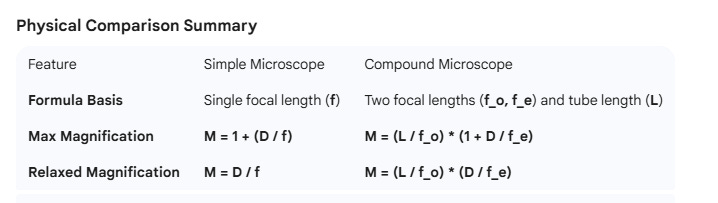
Simple Microscope Magnification:
Maximum (Strain): M = 1 + (D / f)
Minimum (Relaxed): M = D / f
Compound Microscope Magnification:
Total Magnification: M_total = (L / fo) * (D / f_e) (Where L is tube length, fo is objective focal length, and fe is eyepiece focal length)
Quick Tip for NEET Aspirants:
If the question mentions “Normal Adjustment,” always assume the Relaxed Eye (image at infinity).
If the question mentions “Maximum Magnification,” always assume the Eye under Maximum Strain (image at D = 25 cm).
4. Optical Instrument Adjustments: Understanding Normal Adjustment and Peak Magnification
In the context of optical instruments, the “adjustment” refers to the process of changing the distance between the lenses (the objective and the eyepiece) or the distance between the object and the lens to control where the final image is formed relative to the observer’s eye.
Here is a breakdown of the specific adjustments :
a. Normal Adjustment (Relaxed Eye)
This adjustment is used for comfortable, long-duration viewing.
The Goal: To position the intermediate image exactly at the focal point of the eyepiece.
The Mechanism: By adjusting the tube length or the object position, the real image formed by the objective lens is made to fall on the focal point (fe) of the eyepiece.
The Result: Since the “object” for the eyepiece is at its focus, the light rays emerge parallel, and the final virtual image is formed at infinity (∞).
b. Adjustment for Maximum Magnification (Maximum Strain)
This adjustment is used when you need to see the highest level of detail, despite the physical strain on the eye.
The Goal: To position the intermediate image inside the focal length of the eyepiece.
The Mechanism: The lenses are moved closer together (or the object is moved) so that the intermediate image falls between the pole (P) and the focal point (F) of the eyepiece.
The Result: This produces a highly magnified virtual image located exactly at the Near Point (D = 25 cm).
Summary of Adjustment Parameters
Key Physics Logic: In a compound microscope, the objective lens always creates a real, inverted image. The “adjustment” simply shifts where that real image sits in relation to the eyepiece, which then determines if your eye sees a relaxed image at infinity or a strained image at 25 cm.
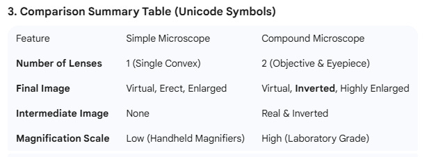
5. The Simple Magnifier (Convex Lens)
A simple microscope uses a single convex lens to solve the “Focus vs. Size” conflict.
Mechanism: The object is placed inside the focal length (u < f).
Result: The lens creates a Virtual, Erect, and Enlarged image.
Win-Win: The image is much larger (large θ) and is formed at or beyond 25 cm, so the eye can focus on it clearly.
6. The Compound Microscope
When a single lens isn’t enough (e.g., for blood cells), we use two lenses in series.
Objective Lens: Small focal length (fo). It forms a Real, Inverted, and Enlarged image of the tiny object.
Eyepiece (Ocular): Acts as a simple magnifier. It takes the image from the objective and magnifies it further into a Virtual Final Image.
7. Key Formulas for NEET
A. Angular Magnification (M)
The ratio of the angle subtended by the image (θ’) to the angle subtended by the object at the near point (θ).
M = θ’ / θ
B. Simple Microscope Magnification
When image is at Near Point (Maximum Strain):
M = 1 + (D / f)
When image is at Infinity (Relaxed Eye):
M = D / f
C. Compound Microscope Magnification
The total magnification is the product of the magnification of the objective (m_o) and the eyepiece (m_e).
Mtotal = (L / f_o) * (D / f_e)
Formula Components:
L: Tube length (distance between the two lenses).
f_o: Focal length of the objective lens.
f_e: Focal length of the eyepiece lens.
D: Least distance of distinct vision ($25 cm$).
8. Critical Reasoning Checklist
Inverted vs. Erect: The final image in a compound microscope is inverted relative to the original object.
Focal Lengths: For high magnification in a microscope, both f_o and f_e should be small (since they are in the denominator).
Relaxed Eye: Always implies the final image is formed at infinity.
10 Angular Magnification: Simple vs. Compound Microscope
The primary goal of any microscope is to increase the visual angle subtended at the eye, which allows the brain to resolve finer details that would otherwise appear as a single blur.
1. Simple Microscope (Single-Stage Magnification)
A simple microscope uses a single convex lens of short focal length. It works by allowing you to place an object very close to your eye—much closer than the standard Near Point (D = 25 cm)—while using the lens to keep the image in focus.
Mechanism: The object is placed inside the focal length (u < f) of the lens.
Image Type: It creates a single Virtual, Erect, and Enlarged image.
Magnification Range: Typically low (usually less than 20x) because very short focal lengths cause image distortion.
2. Compound Microscope (Two-Stage Magnification)
A compound microscope uses two separate convex lenses—the Objective and the Eyepiece—to multiply the magnification effect.
Stage 1 (Objective): This lens is placed near the object and forms a Real, Inverted, and Enlarged image.
Stage 2 (Eyepiece): This lens acts as a simple magnifier. it takes the image from the objective and magnifies it again to create a final Virtual and Inverted image.
Advantage: Because it uses two stages of magnification, it can reach much higher levels (often 1000x or more), allowing us to see microscopic details like bacteria or blood cells.
Why a Compound Microscope is Superior for “Angular Magnification”
The fundamental advantage lies in Multi-Stage Angular Expansion. A Simple Microscope is limited by the physical properties of a single piece of glass, whereas a Compound Microscope uses a two-step process to “stretch” the image across your retina.
1. The Focal Length Constraint (The “f” Barrier)
For a Simple Microscope, the angular magnification is: M = D / f (for a relaxed eye)
To get a very high angular magnification (like 100x), the focal length (f) of the lens would need to be extremely small (0.25 cm).
The Problem: Creating a lens with such a steep curve causes Spherical Aberration (blurring) and Chromatic Aberration (color bleeding).
The Result: The image becomes a “large blurry mess.” You have the “size,” but you lose the “resolution.”
2. The Two-Stage Advantage (Product of Angles)
A Compound Microscope solves this by splitting the task between two lenses. It doesn’t just magnify the object; it magnifies an already magnified image.
Stage 1: The Objective Lens (f_o) It is placed near the object and forms a Real, Inverted, and Enlarged intermediate image.
Stage 2: The Eyepiece (f_e) It acts as a second magnifier. It takes the real image from the objective and increases its Visual Angle (θ′) even further.
The Physics Logic: The total Angular Magnification (M_total) is the product of the two stages: M_total = m_objective × M_eyepiece
If the objective expands the image 40 times and the eyepiece expands that angle another 10 times, the total angular magnification is 400x. This is achieved using two moderately curved lenses, which avoids the distortion of one super-curved lens.
3. Increased Working Distance & Comfort
Simple Microscope: To get high M, you must stick your eye almost touching the lens and the object. This blocks light and causes immense eye strain.
Compound Microscope: The Tube Length (L) separates the lenses, allowing the observer to look through the eyepiece at a comfortable distance while the objective stays close to the specimen.
4. Essential Formulas
Simple Microscope Angular Magnification (M):
Relaxed Eye (Image at ∞): M = D / f
Maximum Strain (Image at 25 cm): M = 1 + (D / f)
Compound Microscope Angular Magnification (M_total): The total magnification is the product of the two lenses.
Combined Formula: Mtotal = (L / f_o) * (D / fe)
Formula Components:
L: Tube length (Distance between lenses).
f_o: Focal length of the objective lens.
f_e: Focal length of the eyepiece lens.
D: Least distance of distinct vision (25 cm).
9. Practice MCQs (NEET Level)
The clarity of an image on the retina is primarily determined by:
A) The brightness of the object
B) The angle subtended at the eye
C) The distance of the ciliary muscles
D) The color of the object
A person cannot see objects clearly closer than 50 cm. To read a book at 25 cm, they need:
A) Concave lens
B) Convex lens
C) Plane mirror
D) Bifocal lens
For a relaxed young eye, the object should be placed at:
A) 10 cm
B) Infinity
C) 25 cm
D) 5 cm
In a simple magnifier, the object is placed:
A) At Focus
B) Between Focus and Center of Curvature
C) Between Pole and Focus
D) At Infinity
The magnification of a simple microscope is 6. If the image is at infinity, the focal length is:
(Take D = 24 cm for easy calculation)
A) 4 cm
B) 6 cm
C) 12 cm
D) 2 cm
In a compound microscope, the intermediate image is:
A) Virtual, erect, magnified
B) Real, inverted, magnified
C) Real, erect, diminished
D) Virtual, inverted, magnified
To increase the magnifying power of a compound microscope:
A) Increase f_o and f_e
B) Decrease f_o and f_e
C) Increase f_o and decrease f_e
D) Decrease f_o and increase f_e
If the focal length of the objective is increased, the magnifying power of the microscope:
A) Increases
B) Decreases
C) Remains same
D) Becomes zero
The tube length of a microscope is the distance between:
A) Object and Objective
B) Objective and Eyepiece
C) Eyepiece and Eye
D) Object and Eye
The final image produced by a compound microscope is:
A) Virtual and erect
B) Real and inverted
C) Virtual and inverted
D) Real and erect
(Questions 11-20 follow similar logic regarding formula application and ray optics properties.)
Answers for Self-Check:
1(B), 2(B), 3(C), 4(C), 5(A), 6(B), 7(B), 8(B), 9(B), 10(C).
NEET Physics: Angular Magnification & Optical Instruments Practice Set
A student views a tiny insect through a simple magnifier. If the insect is moved from 25 cm to 10 cm from the naked eye, the visual angle increases, but the insect looks blurry. Why does the magnifier “fix” this?
A) It decreases the physical size of the insect to fit the retina.
B) It increases the focal length of the eye lens automatically.
C) It creates a virtual image at or beyond 25 cm while maintaining a large visual angle.
D) It converts the virtual image into a real image on the retina directly. (Hint: Think about the “Focus vs. Size” conflict—the eye needs a large angle but also a minimum distance to focus.)
In the formula M = θ′ / θ, what exactly does θ (Theta) represent for a microscope?
A) The angle subtended by the object at the focus of the objective lens.
B) The angle subtended by the object at the near point (25 cm) when viewed by the naked eye.
C) The angle subtended by the image at the infinity point.
D) The angle subtended by the objective lens at the eyepiece. (Hint: θ is the “natural limit” baseline for comparison.)
Two points are so close that they fall on the same photoreceptor cell of the retina. To resolve them, an optical instrument must:
A) Increase the intensity of light hitting that specific cell.
B) Increase the visual angle so the points hit different photoreceptors.
C) Move the points physically further apart on the object.
D) Reduce the tube length of the microscope. (Hint: Think of the retina as pixels; you need the image to cover more “pixels” to see detail.)
For a compound microscope, which state of the eye provides the “Maximum Magnifying Power”?
A) Normal adjustment with the image at infinity.
B) When the ciliary muscles are fully relaxed.
C) When the final image is formed at the Least Distance of Distinct Vision (D = 25 cm).
D) When the intermediate image is at the focal point of the objective. (Hint: Maximum magnification occurs when you “push” the eye to its closest focus limit.)
If you are told a compound microscope is in “Normal Adjustment,” where is the intermediate image located?
A) Exactly at the focal point of the eyepiece (f_e).
B) Between the pole and the focal point of the eyepiece.
C) At the focal point of the objective lens (f_o).
D) At the Near Point (25 cm). (Hint: Normal adjustment is for a relaxed eye, meaning light must leave the eyepiece in parallel rays.)
Why can a “pea” held at arm’s length cover the “Full Moon” in your field of vision?
A) The linear size of the pea increases as it gets closer.
B) The pea subtends a larger visual angle at the eye than the moon does.
C) The moon’s light rays are parallel while the pea’s rays are converging.
D) The pea has a higher angular magnification than the moon. (Hint: Our brain perceives size based on the angle occupied on the retina, not absolute kilometers.)
To achieve a very high total magnification in a compound microscope, what should be the focal lengths of the objective (f_o) and eyepiece (f_e)?
A) Both f_o and f_e should be very large.
B) f_o should be large and f_e should be small.
C) Both f_o and f_e should be small.
D) f_o should be small and f_e should be large. (Hint: Look at the denominator of the formula: M_total ≈ (L / f_o) × (D / f_e).)
Which of the following describes the nature of the final image formed by a compound microscope?
A) Real, Inverted, and Enlarged.
B) Virtual, Erect, and Enlarged.
C) Virtual, Inverted (relative to the original object), and Enlarged.
D) Real, Erect, and Diminished. (Hint: The objective inverts the image once; the eyepiece magnifies that inversion.)
A simple microscope has a focal length “f”. If the user wants the most comfortable (least strained) view, the magnification M will be:
A) M = 1 + (D / f)
B) M = D / f
C) M = f / D
D) M = (L × D) / (f_o × f_e) (Hint: Relaxed viewing means the image is formed at infinity.)
In a compound microscope, the tube length (L) is essentially the distance between:
A) The object and the objective lens.
B) The eyepiece and the observer’s eye.
C) The focal point of the objective and the focal point of the eyepiece.
D) The eye’s near point and the infinity point. (Hint: L represents the internal optical path between the two lenses.)
What happens to the magnifying power of a simple microscope if it is used underwater (where the focal length “f” of the lens increases)?
A) The magnifying power increases.
B) The magnifying power decreases.
C) The magnifying power remains the same.
D) The magnifying power becomes infinite. (Hint: M = D / f. If the denominator “f” gets bigger, what happens to the result?)
If an instrument has M = 15, what does this tell the “brain” about the object?
A) The object has physically grown 15 cm in height.
B) The object is 15 times closer than the eye’s near point.
C) The object occupies 15 times more space on the retina than it would at 25 cm.
D) The object’s light is 15 times brighter. (Hint: Angular magnification measures perceived space enhancement.)
Which component of a compound microscope acts like a simple magnifier?
A) The objective lens.
B) The tube length adjustment knob.
C) The eyepiece lens.
D) The mirror or light source. (Hint: The first lens creates an image; the second lens magnifies that image for your eye.)
A bacteria is invisible to the naked eye because it subtends an angle θ < 1 minute. A microscope makes θ′ > 1 minute. The result of seeing it clearly is called:
A) Linear magnification.
B) Resolution.
C) Refraction.
D) Ciliary contraction. (Hint: This is the ability to distinguish detail rather than just “blowing up” a blur.)
In a compound microscope, if you move the objective lens slightly further from the object (increasing u), the intermediate image will move:
A) Closer to the eyepiece.
B) Closer to the objective lens.
C) To the Near Point (D).
D) To infinity. (Hint: Apply the lens formula 1/v - 1/u = 1/f; as object distance increases, image distance decreases.)
Answer Key & Brief Rationales:
C - It solves the “Focus vs. Size” conflict by placing a large-angled image at a focusable distance.
B - Theta is always the “Naked Eye” reference angle at D = 25 cm.
B - Increasing the angle spreads light across more photoreceptors (pixels).
C - Image at D (25 cm) provides the largest angle, though it causes eye strain.
A - Parallel rays for a relaxed eye are only possible if the intermediate image is at the eyepiece focus.
B - Perceived size is determined by the Visual Angle (θ).
C - Both f_o and f_e are in the denominator; smaller values increase the total M.
C - Virtual (eyepiece effect) and Inverted (objective effect).
B - The formula for infinity (relaxed) is D/f.
C - Tube length L is the distance between the focal points of the two lenses.
B - Larger focal length means lower magnifying power (M ∝ 1/f).
C - M = 15 means it takes up 15 times the “retinal real estate.”
C - The eyepiece magnifies the intermediate real image.
B - Resolution is the ability to distinguish points as separate.
B - Increasing object distance (u) results in a smaller image distance (v).
NEET Conceptual Challenge: Assertion & Reason
Instructions:
A: Both Assertion and Reason are true, and Reason is the correct explanation of Assertion.
B: Both Assertion and Reason are true, but Reason is NOT the correct explanation of Assertion.
C: Assertion is true, but Reason is false.
D: Both Assertion and Reason are false.
Assertion (A): In a Compound Microscope, the total angular magnification is high because the objective lens produces a virtual and enlarged intermediate image which is then further magnified by the eyepiece.
Reason (R): A Simple Microscope provides lower angular magnification because it decreases the visual angle (θ) of the object compared to the angle subtended at the near point (25 cm) to keep the image in focus.
Correct Answer: D (Both are False)
Why the Assertion (A) is False:
The Assertion sounds correct because a Compound Microscope does have high magnification. However, the “trap” is the word virtual.
In reality, the objective lens forms a Real and Inverted intermediate image.
If the intermediate image were virtual, the eyepiece (which acts as a simple magnifier) would not be able to “pick it up” and magnify it further in the standard two-stage configuration.
Why the Reason (R) is False:
The Reason sounds logical because simple microscopes have limits, but the physics is reversed.
A Simple Microscope increases the visual angle (θ′ > θ).
The Reason claims it “decreases” the angle. If an instrument decreased the visual angle, it would be a “minifier” (making things look smaller), not a microscope.
The actual purpose of the lens is to allow the object to be brought closer than 25 cm (to increase the angle) while using the lens to fix the focus.
Student Feedback Loop:
The “A” Trap: Students who focus only on the word “High Magnification” will think A is true.
The “R” Trap: Students who confuse “keeping the image in focus” with “reducing the angle” will fall for R.
NEET Conceptual Challenge
Instructions:
(A) Both Assertion and Reason are true, and Reason is the correct explanation of Assertion.
(B) Both Assertion and Reason are true, but Reason is NOT the correct explanation of Assertion.
(C) Assertion is true, but Reason is false.
(D) Both Assertion and Reason are false.
Assertion (A): The primary physical significance of a compound microscope is its ability to increase the visual angle (θ′) of a microscopic object while allowing the final image to be formed at or beyond the Near Point (D = 25 cm).
Reason (R): The human eye cannot resolve the details of a tiny object simply by bringing it very close to the eye, because even though the visual angle (θ) increases, the ciliary muscles cannot increase the power of the eye lens enough to focus the image on the retina.
Correct Answer: (A)
1. Why the Assertion is True: The “Magnifying Power” of a microscope is essentially Angular Magnification. It isn’t just about linear size; it’s about “retinal real estate.” By increasing the angle θ′, the microscope ensures the image covers more photoreceptors. Crucially, it must place that image at least 25 cm away (the Near Point) or at Infinity so the eye can actually see it clearly.
2. Why the Reason is True: If you take a needle and hold it 2 cm from your eye, the angle it makes is massive! Theoretically, you should see every atom. But you don’t—you see a blur. This is because the eye’s lens has a limit to how much it can bulge (maximum power). The Reason correctly identifies that the biological limit of focus is what makes the microscope’s angular magnification necessary.
3. Why R explains A: The Reason explains the “Why” behind the “How.” We need the microscope to increase the angle (Assertion) because our eyes fail to focus on large angles naturally at close distances (Reason). This “Focus-Size” conflict is the heart of optical instrument physics.
Quick Summary for Students:
The Conflict: Bringing things closer makes them “bigger” (angle increases) but “blurrier” (focus fails).
The Solution: The Compound Microscope uses two stages of magnification to give you the Large Angle of a close object with the Perfect Focus of a distant one.